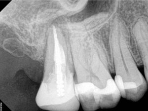
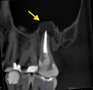
Root canals are often diagnosed using cone beam computed tomography. Advanced Endodontics uses cone beam computed tomography (CBCT). CBCT is a 3D imaging modality used to best diagnose and treat a tooth. It uses the principles from medical CT imaging to scan the specific teeth in question. It is a twenty-second scan. The sensor moves around your head like panoramic radiographs utilized by most dental offices. The 3D image gives us tremendous detail and information. This information helps us most accurately determine which teeth are affected and why, and then allows us work very precisely and conserve as much tooth structure as possible during your treatment.
Root Canal Diagnostics Enhanced
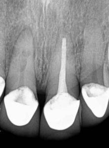
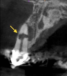
The 3D scan provides much more information than the traditional 2D images. We are able to more accurately diagnose diseased teeth and any affected surrounding areas. Numerous studies have demonstrated the CBCT to be much more accurate than traditional radiography in identifying diseased teeth1-7. In the example below (Figures 1 and 2), the patient was referred by his ear, nose and throat surgeon after having a long history of sinus congestion and nasal discharge with no discernible reason. The 2D image is unclear, but the 3D image clearly shows a large area of inflammation around the tooth that is invading the sinus. This tooth was treated three years ago, and the patient still has the tooth and no more symptoms or sinus issues.
Precise Root Canal Plan & Treatment
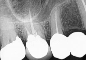
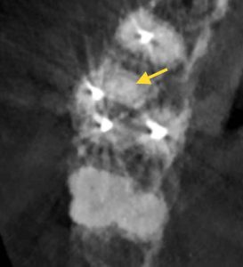
With all of the extra information we get from the 3D scan, we are able to plan and complete our treatment in a more precise and conservative manner. We know exactly where all of the areas that need treated are, so that we can be confident and preserve as much of the tooth structure as possible. In our second case (Figures 3 and 4), we identified a canal that was missed when her initial root canal was done that had become infected. We would not have been able to plan the treatment as conservatively with just the 2D images. In our third case (Figures 5 and 6), the tooth had extensive disease. The location and extent is much clearer on the 3D scan, which aided our decision making in determining the best plan to treat the disease and save his front tooth. Several studies have demonstrated that 3D imaging frequently influences providers’ decisions of how best to treat the tooth because of the greater amount of knowledge they have about the situation 8-10.
Root Canal Safety
Everyone’s concern when it comes to radiography is safety. And for good reason! Ionizing radiation can be harmful in certain types and doses. Fortunately, medical and dental radiation are very low-dose, low-risk, and the information gained is incredibly beneficial. The American Association of Physicists in Medicine found inconclusive evidence of any harmful effects from a medical imaging dose below 100,000 μSv11. The Endodontic CBCT dose is typically around 20-40 μSv12. Far below that threshold! Even for pregnant patients, who are often considered to be greater risk, the American College of Obstetricians and Gynecologists Committee on Health Care for Underserved Women13 has stated: “…prevention, diagnosis, and treatment of oral conditions, including dental X-rays (with shielding of the abdomen and thyroid) … [is] safe during pregnancy.” We use the most up to date equipment and recommended techniques for shielding to keep you and our team safe while obtaining the most accurate and comprehensive information we can to best take care of you.
Conclusion
A brief twenty second scan is all the time needed to provide you safe, comfortable and accurate care. We are happy to review your scan with you to show you all of the information we have gained and describe how we will use to to take care of you and your tooth.
References:
- Paula-Silva FWG, Wu MK, Leonardo MR, da Silva LAD, Wesselink PR Accuracy of periapical radiography and cone-beam computed tomography scans in diagnosing apical periodontitis using histo-pathological findings as a gold standard. J Endod 2009;35:1009–12.
- Paula-Silva FWG, Junior MS, Leonardo MR, Consolaro A, Silva LAB, Preto R (2009b) Cone- beam computerized tomographic, radiographic, and histological evaluation of periapical repair in dogs post-endodontic treatment. Oral Surgery, Oral Medicine, Oral Pathology, Oral Radiology, and Endodontology 108, 796–805.
- Lofthag-Hansen S, Huumonen S, Gro€ndahl K, Gro€ndahl H-G (2007) Limited cone-beam CT and intraoral radiography for the diagnosis of periapical pathology. Oral Surgery, Oral Medicine, Oral Pathology, Oral Radiology, and Endodontology 103, 114-9
- Low MTL, Dula KD, Bu€rgin W, von Arx T Comparison of periapical radiography and limited cone-beam tomography in posterior maxillary teeth referred for apical surgery. J Endod 2008;34:557–62.
- Bornstein MM, Lauber R, Sendi P, von Arx T (2011) Com- parison of periapical and limited cone-beam computed tomography in mandibular molars for analysis of anatomical landmarks before apical surgery. Journal of Endodontics 37, 151–7.
- Patel S, Wilson R, Dawood A, Mannocci F (2012a) The detection of periapical pathosis using periapical radiography and cone beam computed tomography – Part 1: pre-operative status. International Endodontic Journal 45, 702–10.
- Patel S, Wilson R, Dawood A, Foschi Mannocci F (2012b) The detection of periapical pathosis using digital periapical radiography and cone beam computed tomography – Part 2: a 1-year post-treatment follow-up. International Endodontic Journal 45, 711–23.
- Patel K, Mannocci F, Patel S. The assessment and management of external cervicalresorption with periapical radiographs and cone-beam computed tomography. J Endod 2016;42:1435–40.
- Goodell K., Mines P., Kersten D. Impact of Cone-Beam Computed Tomography on Treatment Planning for External Cervical Resorption and a Novel Axial Slice-based Classification System J Endod, 2017;44:239–244
- Viana Wanzeler, AM., et al. Can Cone-beam Computed Tomography Change Endodontists’ Level of Confidence in Diagnosis and Treatment Planning? A Before and After Study J Endodon, 2020;46:283-288
- AAPM Position Statement on Radiation Risks from Medical Imaging Procedures PP25-C 2018
- Patel S, Durack C, Abella F, Shemesh H, Roig M, Lemberg K. Cone beam computed tomography in Endodontics – a review. International Endodontic Journal, 48, 3–15, 2015.
- American College of Obstetricians and Gynecologists Committee on Health Care for Underserved Women. Committee Opinion: Oral health care during pregnancy and through the lifespan. American College of Obstetricians and Gynecologists August 2013 (Reaffirmed 2017)
